Welcome to the monthly SHDA Research Update. Our specialists have selected 3 seminal papers that have been recently published in each speciality (perioperative medicine, cardiac imaging, cardiology, cardiothoracic surgery).
Perioperative Medicine
Summarised by Sarah Catchpoole
The authors present an extensive overview and update of right heart imaging in the perioperative setting. Nonstandard and further views are discussed in detail and assessed by a case series of 30 subjects undergoing routine TOE for elective cardiac surgery.
Increasingly complex applications of 3D echocardiography are becoming established in cardiac surgery and in the catheter laboratory. This brief editorial provides an overview of advantages of 3D echocardiography in assessment of volume, length, and area in a range of pathologies and settings. Disadvantages, inaccuracies and practical perioperative considerations are also discussed.
Methodology for the measurement of tricuspid annulus (TA) size and thresholds for TA enlargement are not clearly defined. This prospective study compared 2D-TTE (n = 282), 3D-TOE (n = 183), and 2D-TTE in healthy controls (n= 66) with surgical measurement (n = 120) of TA diameter. 2D-TTE measurements in the apical 4 chamber position offered the highest accuracy, feasibility and reproducibility, but were systematically smaller than the LA diameter of the TA measured using 3D-TEE. The authors also found surgical measurements were poorly reproducible as a slight traction on the TA resulted in marked changes in TA measurements.
Cardiac Imaging
Summarised by Sarah Catchpoole
The correlation between measurements of pulmonary artery pressure by TTE and right heart catheterization (RHC) are debated. While RHC is the gold standard, TTE is far more desirable due to its non-invasive approach. In a prospective cohort of 300 patients with significant rheumatic mitral stenosis, the authors found good correlation between systolic and mean pulmonary artery pressures (sPAP and mPAP) measured by RHC and TTE (p < 0.001 for all comparisons). Sensitivity and specificity were acceptable at 92.8% and 86.6% respectively for TTE measured sPAP, and 94.1% and 73.3% respectively for TTE measured mPAP.
As transcatheter aortic valve replacement (TAVR) is becoming more common, so too is the number of patients requiring follow-up increasing. Grant et al. provide a brief overview for clinicians of the haemodynamic and echocardiographic parameters to monitor for stenosis, para-prosthetic aortic valve regurgitation, and prosthesis–patient mismatch.
The mainstay of initial assessment of aortic stenosis remains echocardiography, but complementary imaging methods such as aortic valve CT calcium scoring, PET/CT, and cardiovascular magnetic resonance have prognostic roles. This comprehensive review provides a guide to clinicians on current imaging assessment of aortic stenosis, with consideration to disease progression.
Cardiology
Summarised by Sarah Catchpoole
The introduction of new mitral valve therapies such as transcatheter devices has highlighted the need for a consensus approach to pragmatic clinical trial design. This document, the first of two consensus statements from the international Mitral Valve Academic Research Consortium, provides guiding principles as a template for clinical investigation of mitral therapeutics. The report includes discussion of classification, grading, quantification, and the role of novel imaging.
In symptomatic patients with normal flow, low-gradient severe aortic stenosis (AS) and preserved LVEF, timing of aortic valve replacement (AVR) remains controversial. In a prospective consecutive cohort of 284 patients, Kang et al. found no significant differences between the early AVR (n = 98) and watchful observation groups (n = 186) for the risk of overall mortality, the estimated actuarial 8- year mortality rates, or risk of overall death in propensity-score-matched pairs (n = 88). Therefore, they conclude watchful observation with timely performance of AVR should be considered a therapeutic option for low-gradient severe AS.
Basso et al. Arrhythmic Mitral Valve Prolapse and Sudden Cardiac Death. Circulation. 2015 Aug
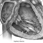
MVP in young adults may present with ventricular arrhythmias and sudden cardiac death (SCD), however the pathogenesis of this and risk stratification remains controversial. Basso et al. found left ventricular late enhancement (identified by contrast-enhanced cardiac magnetic resonance) in 93% of of MVP patients vs. 14% of healthy control subjects (p < 0.001). The authors also investigated the LV myocardium to search for the substrate of electric instability for the first time. Left ventricular fibrosis was detected on histology at the level of papillary muscles in all patients, and inferobasal wall in 88%. Therefore, fibrosis (suggesting a myocardial stretch by the prolapsing leaflet) is the structural hallmark and correlates with ventricular arrhythmia origin.
Cardiothoracic Surgery
Summarised by Andrew Haymet
Mitroflow pericardial valves are an ideal aortic valve prosthesis for patients with a small aortic annulus. The structural performance of 28 valves implanted for a 12 year period was analyzed by gross and microscopic examination after explantation. The mean age of implantation was 72.1 ± 5.69 years, with an interval between index surgery and explantation of 4.5 ± 3.4 years. Structural deterioration was observed in 18 of 19 valves which had been implanted for at least 30 months. Modes of failure included cusp thickening, tears, pannus deposition and calcification. Given the early deterioration demonstrated by Mitroflow valves, their use should be carefully assessed in all eligible patients, including those aged over 65 years.
In this propensity matched study mitral valve surgery through a right minithoracotomy (RT) was compared to the standard approach through median sternotomy (215 patients in each group). The RT approach was associated lower rates of new-onset atrial fibrillation (8% vs 16%; p=0.018), pneumonia (1% vs 5%; p=0.049), respiratory failure (3% vs 8%; p=0.036), and acute renal failure (2% vs 7%; p=0.006); lower chest tube output (350 vs 840mL; p<0.001), and fewer red blood cell transfusions (2 vs 3 units; p=0.001). Based on this study at a single institution, the RT approach may confer some benefit over median sternotomy for selected patients.
 The Cavalier trial evaluated the Perceval sutureless valve in 658 consecutive patients in 25 European centres between 2010 to 2013. Successful implantation occurred in 95.4% of patients. The 30 day overall and valve related mortality was 3.7% and 0.5%, respectively. The mean effective orifice area improved from 0.72 to 1.46cm2. Mean cardiopulmonary bypass & cross clamp time when the valve was used in isolated AVR through a median sternotomy was 53.4 ± 20.5 & 32.4 ± 10.9, respectively. The authors found using the Perceval valve a safe and reproducible procedure, and providing good early clinical outcomes.


