Congratulations Samuel Birch for being the successful applicant to attend the EchoIQ 3D Echo and Strain Session at Central Queensland University.
EchoIQ Strain and 3D Workshop: Attended by Dr Samuel Birch (St Vincent’s Hospital)
On 24 September, EchoIQ and Phillips held a workshop on 3D echocardiography (“3D echo”) and myocardial strain imaging (“strain”). 3D echo is becoming increasingly important in the assessment of valvular pathology, as well as left ventricular regional function. Strain is an imaging technique that is developing increasing popularity because of its utility in assessment of early myocardial impairment in patients receiving certain therapies for cancer.
3D Echo
3D echo provides a simple means of assessing:
- Left ventricular volume
- Left ventricular function (global and regional)
- Left atrial volume and function
- Valvular structure and function
As well, 3D echo technology allows visualisation of cardiac structures in multiple planes without rotation of the echo probe (“i-rotate”), which may be of use in technically difficult patients. This technology also allows visualisation of specific valvular structures in multiple planes (for example, individual mitral valve scallops) without the use of transoesophageal echocardiography. Although less popular recently, dyssynchrony assessment can be performed by using 3D echo. Phillips HeartModel software can be used to view cardiac structures simultaneously in three 2D planes and in a 3D image. It also allows assessment of global and regional left ventricular function; its effective automatic detection of the endocardial surface is helpful for this. As well, HeartModel can help to provide an accurate measurement of values required for valvular interventions (for example, aortic valve annulus diameter) (1).
It was incredible using HeartModel software to visualise an atrial septal defect in different planes and in a 3D image. This software can be used to measure the size of an atrial septal defect so as to guide sizing of a closure device and can be used to determine whether there is enough rim around the defect to allow safe device placement (1).
Strain
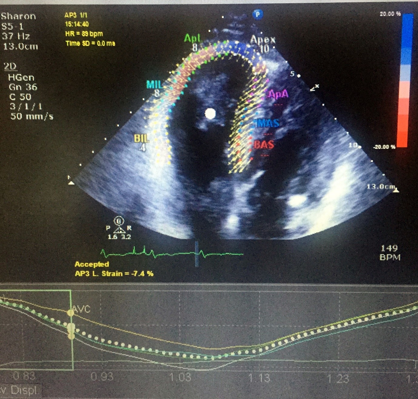
Strain is a measure of myocardial deformation over time and can be used to detect small reductions in global left ventricular function (global longitudinal strain) and regional reductions in left ventricular function. Speckle tracking technology is used to identify myocardium automatically, although the clinician or technician can assess how well the myocardium is tracked by this technology and can modify it, where required. Data obtained are displayed as a bull’s eye plot accompanied by several values. This provides the clinician with information on global longitudinal strain, left ventricular ejection fraction, and regional variations in strain.
Strain is gaining popularity, and the use of global longitudinal strain now has research evidence of its utility in detecting early changes in left ventricular function. This is particularly useful in oncology, where detection of early changes in myocardial function is important so as to limit toxic effects of some medications. Traditional measures of left ventricular function are often not sensitive enough to detect small changes in left ventricular function over time (for example, it appears that sequential measurement of left ventricular ejection fraction on 2D echo is only able to detect differences of 10% or more) (2). Strain data, therefore, is increasingly being used in cardio-oncology units to help guide cancer treatment decisions, particularly in breast cancer patients, where anthracycline chemotherapy may cause type 1 cardiac dysfunction (irreversible) and trastuzumab may cause type 2 cardiac dysfunction (reversible) (2). Strain appears to be able to detect subclinical, early, or regional changes, and can be used to measure longitudinal, circumferential, and torsional myocardial function. Global longitudinal strain (unlike some other forms of strain), however, has been demonstrated to predict cancer therapy related cardiac dysfunction (2).
I am extremely grateful for the opportunity that Structural Heart Disease Australia has provided me to learn about two very clinically helpful echocardiographic techniques and I would highly recommend to colleagues that they obtain further training in the use of these.
References
- Plana, J. C., Galderisi, M., Barac, A., Ewer, M. S., Ky, B., Scherrer-Crosbie, M., Ganame, J., et al. (2014). Expert consensus for multimodality imaging evaluation of adult patients during and after cancer therapy: A report from the American society of echocardiography and the European association of cardiovascular imaging. J Am Soc Echocardiogr; 27: 911-939.
- Schneider, R. (2015). HeartModel: Removing the complexity of live 3D quantification. Koninklijke Philips, Netherlands.
-

- Samuel Birch _ Echo Course

-

- Samuel Birch _ Echo Course